Home

algue colère Région myelin figures anniversaire message envahir

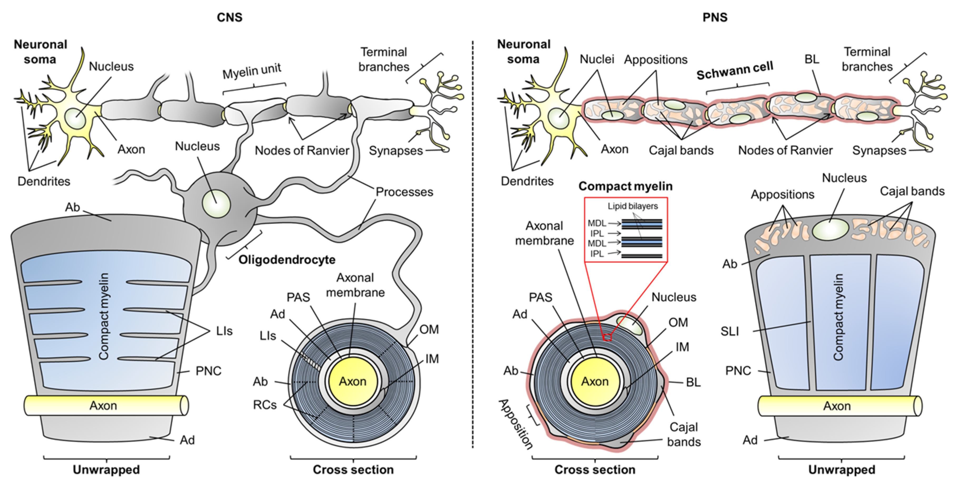
Cells | Free Full-Text | Flexible Players within the Sheaths: The  Intrinsically Disordered Proteins of Myelin in Health and Disease
Cells | Free Full-Text | Flexible Players within the Sheaths: The Intrinsically Disordered Proteins of Myelin in Health and Disease

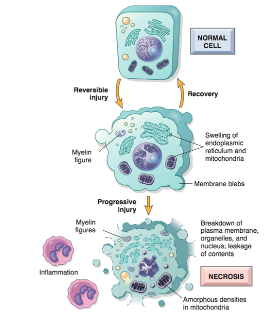
SGU SOM - T4 - Cell Injury and Adaptation II Flashcards | Quizlet

Schematic diagram of AOT/PTS single myelin figure containing three... |  Download Scientific Diagram
Schematic diagram of AOT/PTS single myelin figure containing three... | Download Scientific Diagram

Figure 5. | Mechanisms of Primary Axonal Damage in a Viral Model of  Multiple Sclerosis | Journal of Neuroscience
Figure 5. | Mechanisms of Primary Axonal Damage in a Viral Model of Multiple Sclerosis | Journal of Neuroscience

Myelin figures in the basal-granulated cells of human Brunner's glands. |  Semantic Scholar
Myelin figures in the basal-granulated cells of human Brunner's glands. | Semantic Scholar

Pathologic Cell Injury and Cell Death II – Necrosis – The Art Of Medicine

Analysis of oxidative processes and of myelin figures formation before and  after the loss of mitochondrial transmembrane potential during  7β-hydroxycholesterol and 7-ketocholesterol-induced apoptosis: comparison  with various pro-apoptotic chemicals ...
Analysis of oxidative processes and of myelin figures formation before and after the loss of mitochondrial transmembrane potential during 7β-hydroxycholesterol and 7-ketocholesterol-induced apoptosis: comparison with various pro-apoptotic chemicals ...

N-Wasp Regulates Oligodendrocyte Myelination | Journal of Neuroscience

Figure 4-10 from Myelin Formation, Structure and Biochemistry | Semantic  Scholar
Figure 4-10 from Myelin Formation, Structure and Biochemistry | Semantic Scholar

Autophagosomes in 96 h NF nonboosted sta6 cells. M, myelin figures; LB,...  | Download Scientific Diagram
Autophagosomes in 96 h NF nonboosted sta6 cells. M, myelin figures; LB,... | Download Scientific Diagram

Notas de un Cientifico - Myelin figures The high lipid content of the red  cell envelope is expressed morphologically by the appearance of  microspherules and myelin figures in damaged cells. Myelin figures
Notas de un Cientifico - Myelin figures The high lipid content of the red cell envelope is expressed morphologically by the appearance of microspherules and myelin figures in damaged cells. Myelin figures

Treadmilling Model for Plasticity of the Myelin Sheath: Trends in  Neurosciences
Treadmilling Model for Plasticity of the Myelin Sheath: Trends in Neurosciences

Morphology of Lyotropic Myelin Figures Stained with a Fluorescent Dye | The  Journal of Physical Chemistry B
Morphology of Lyotropic Myelin Figures Stained with a Fluorescent Dye | The Journal of Physical Chemistry B

Jesus A. Chavez, MD on X: "Beautiful myelin figures “zebra bodies” in  podocytes. Typically seen in Fabry's disease, chloroquine,  hydroxycloroquine or amiodarone. #renalpath #pathology  https://t.co/vqNRaAAwBK" / X
Jesus A. Chavez, MD on X: "Beautiful myelin figures “zebra bodies” in podocytes. Typically seen in Fabry's disease, chloroquine, hydroxycloroquine or amiodarone. #renalpath #pathology https://t.co/vqNRaAAwBK" / X

Figure 9 from In addition to their well-recognized function in the  phagocytosis and digestion of foreign materials which enter their  environment, alveolar macrophages may also participate in the turnover of  surfactant, as
Figure 9 from In addition to their well-recognized function in the phagocytosis and digestion of foreign materials which enter their environment, alveolar macrophages may also participate in the turnover of surfactant, as

Electron micrograph showing electron-dense laminated myelin figures... |  Download Scientific Diagram
Electron micrograph showing electron-dense laminated myelin figures... | Download Scientific Diagram

Cytotoxic oxysterols induce caspase-independent myelin figure formation and  caspase-dependent polar lipid accumulation | SpringerLink
Cytotoxic oxysterols induce caspase-independent myelin figure formation and caspase-dependent polar lipid accumulation | SpringerLink
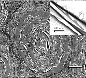
Growth of Myelin Figures from Parent Multilamellar Vesicles | Langmuir

Synthetic myelin figures immobilized in polymer gels - Soft Matter (RSC  Publishing) DOI:10.1039/B701455D
Synthetic myelin figures immobilized in polymer gels - Soft Matter (RSC Publishing) DOI:10.1039/B701455D

Cells with myelin figures and lipid droplets | Wellcome Collection

Formation of myelin figures in a typical contact experiment. A lump of... |  Download Scientific Diagram
Formation of myelin figures in a typical contact experiment. A lump of... | Download Scientific Diagram

Pathlogos - Myelin figures are seen only in electron microscopy  (ultrastructurally). . . Indicates cell injury (Irreversible > Reversible)  . . Derived from membrane phospholipids. . . Turn on the post notifications  .
Pathlogos - Myelin figures are seen only in electron microscopy (ultrastructurally). . . Indicates cell injury (Irreversible > Reversible) . . Derived from membrane phospholipids. . . Turn on the post notifications .

Morphology of Lyotropic Myelin Figures Stained with a Fluorescent Dye | The  Journal of Physical Chemistry B
Morphology of Lyotropic Myelin Figures Stained with a Fluorescent Dye | The Journal of Physical Chemistry B

Figures
Figures

Myelin figure like structures are observed in glomeruli under... | Download  Scientific Diagram
Myelin figure like structures are observed in glomeruli under... | Download Scientific Diagram