Home

Ruiné Artisanat Tragique hypodense et hyperdense scanner cassé dinosaure béton

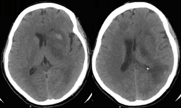
Acute bleed is hyperdense and chronic is hypodense. | Download Scientific  Diagram
Acute bleed is hyperdense and chronic is hypodense. | Download Scientific Diagram

Radiologie et imagerie medicale - difference entre HSD et HED | Facebook

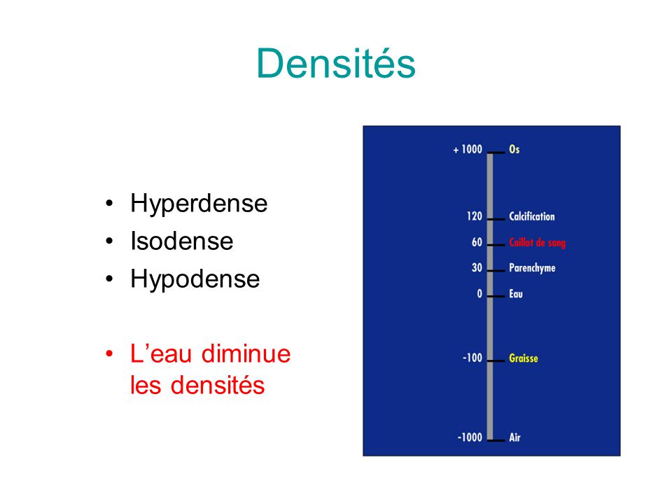
Neuroradiologie - Neuroimagerie - ppt video online télécharger

Semiologie semio neuroradio | PPT

PPT - Neuroradiologie - Neuroimagerie PowerPoint Presentation, free  download - ID:3526927
PPT - Neuroradiologie - Neuroimagerie PowerPoint Presentation, free download - ID:3526927

Apport de la Tomodensitométrie dans les Pathologies Infectieuses  Cérébroméningées au CHU du Point G
Apport de la Tomodensitométrie dans les Pathologies Infectieuses Cérébroméningées au CHU du Point G

Scanner (CT)
Scanner (CT)

hypodense and hyperdense

Examples of the four types of hyperdense lesions on the non-contrast CT...  | Download Scientific Diagram
Examples of the four types of hyperdense lesions on the non-contrast CT... | Download Scientific Diagram

Apport de l'imagerie neurologique dans la prise en charge du patient  traumatisé crânien grave
Apport de l'imagerie neurologique dans la prise en charge du patient traumatisé crânien grave

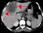
John Libbey Eurotext - Hépato-Gastro & Oncologie Digestive -  Tomodensitométrie hépatique II. Foie pathologique : pathologie tumorale
John Libbey Eurotext - Hépato-Gastro & Oncologie Digestive - Tomodensitométrie hépatique II. Foie pathologique : pathologie tumorale
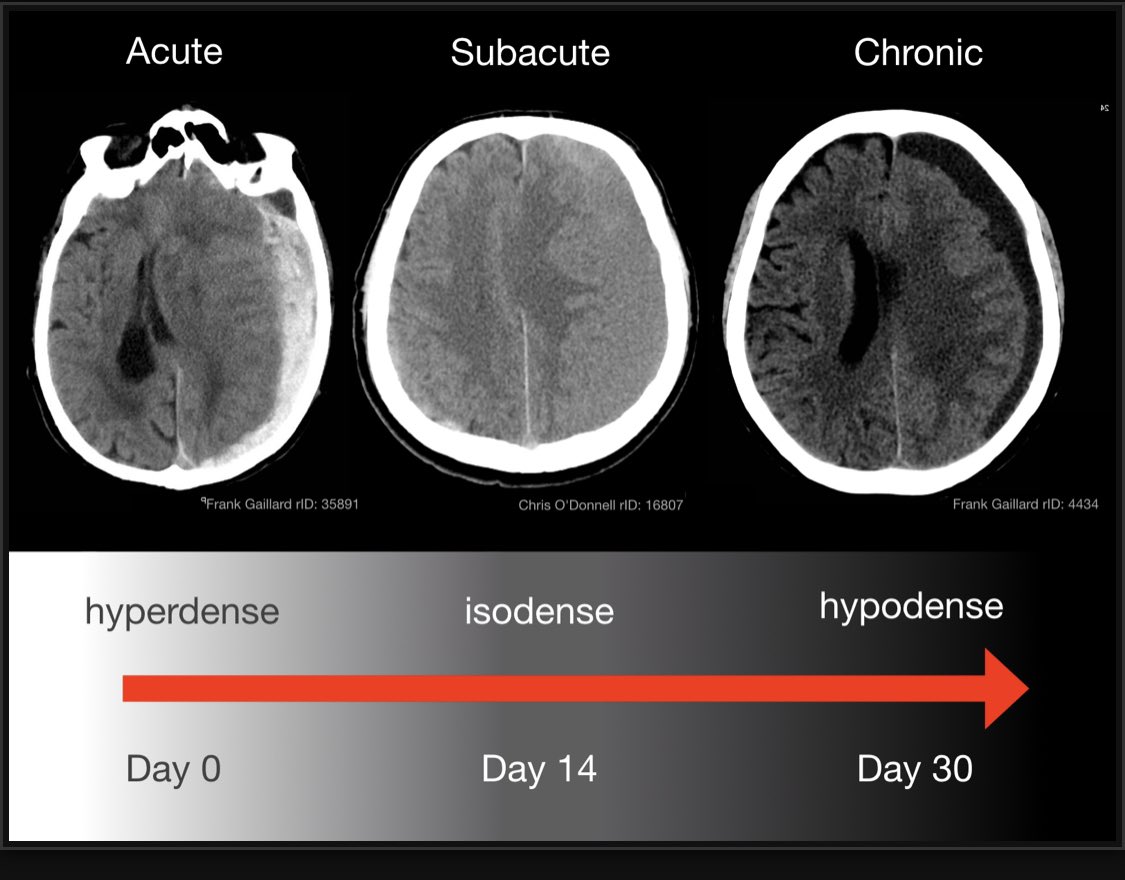
Nitin Arora on X: "The appearance of blood in CT scan changes with time  #Radmasterclass #ICSSOA2018 https://t.co/tEOUe4XdGA" / X
Nitin Arora on X: "The appearance of blood in CT scan changes with time #Radmasterclass #ICSSOA2018 https://t.co/tEOUe4XdGA" / X

Axial T2-weighted MRI of the brain shows a mass with hypodense and... |  Download Scientific Diagram
Axial T2-weighted MRI of the brain shows a mass with hypodense and... | Download Scientific Diagram

Imagerie de l'accident vasculaire cérébral ischémique à la phase aigue  Imaging of Acute Stroke
Imagerie de l'accident vasculaire cérébral ischémique à la phase aigue Imaging of Acute Stroke

How to interpret an unenhanced CT Brain scan. Part 2: Clinical cases

Semiologie semio neuroradio | PPT

Diaporama : le scanner cérébral sans injection | Medscape

Hématome sous-dural
Hématome sous-dural

How can a hypodense area be located in a CT scan? - Quora

Semiologie semio neuroradio | PPT

Diaporama : le scanner cérébral sans injection | Medscape

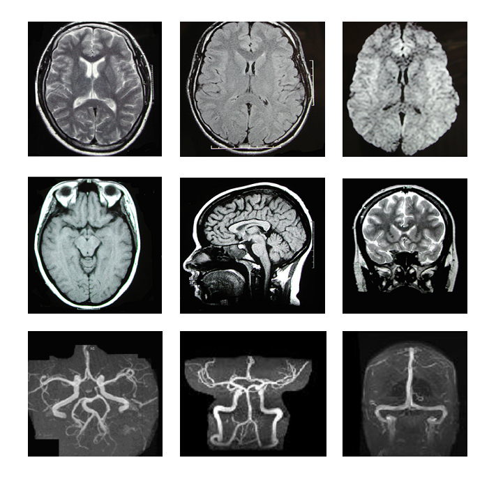
Neuro-imagerie : TDM, IRM et ARM, angiographie - Le livre de sémiologie  médicale
Neuro-imagerie : TDM, IRM et ARM, angiographie - Le livre de sémiologie médicale

Cours n°25 : Sémiologie neuroradiologique

Origine infectieuse des lésions cérébrales à rehaussement annulaire

John Libbey Eurotext - Hépato-Gastro & Oncologie Digestive -  Tomodensitométrie hépatique : 1. les affections non néoplasiques
John Libbey Eurotext - Hépato-Gastro & Oncologie Digestive - Tomodensitométrie hépatique : 1. les affections non néoplasiques